Skin Scanning

Individuals now have access to an affordable and innovative skin scanning service.

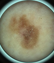
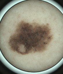
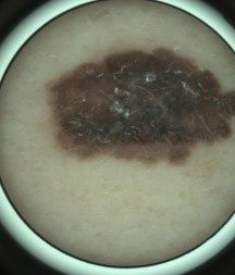
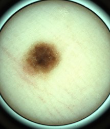
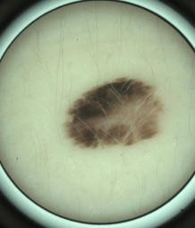
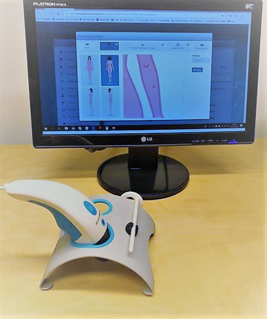
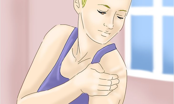
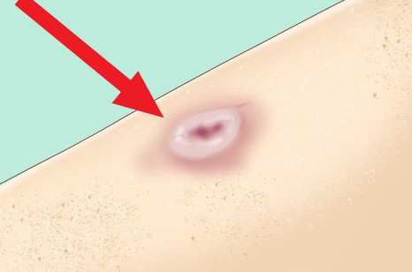
The process is simple and quick. In a consultation clients will be asked some questions about their general skin health including previously used medications etc. The skin condition is imaged with our non-invasive camera, looking at the surrounding skin and a very high-quality close-up image of the area of concern.

The images are sent to a dermatology consultant who analyses the images remotely, along with the supporting information. Once the results are received, we will call to discuss the report and then send it via email. If needed an additional appointment will be made with the Doctor.

Know your ABC’s…

Use the ABCDE rule as a tool to assess characteristics of normal moles vs abnormal moles.

ASYMMETRY
An irregular shape. The 2 halves should be symmetrical.

BORDERS
Look for unclear, irregular, or ragged borders.

COLOUR
Changes in colour. Especially black, blue or uneven colour.

DIAMETER
More than 5 – 6mm in diameter and change in size.

EVOLVING
Changes in shape, size, colour. Itching or bleeding of an existing mole, or if a new mole/lesion appears.

MOLE SCANNING SERVICE

“Early detection improves outcomes and saves lives”

Did you know?

Skin cancer is the most common cancer overall in the UK.
(Cancer Research 2016)

Malignant Melanoma has increased by 400% over the last 35 years and is now the second most common cancer in people between the ages of 20 to 40 years.
(Cancer Research 2016)

90% of all skin cancers are caused by over-exposure to UV radiation from the sun and/or sunbeds – that means the majority of all skin cancers are preventable by undertaking simple sun safety measures.

Individuals now have access to an affordable, innovative mole/pigmented lesion scanning service.

The process is simple and quick. In a consultation the client will be asked some questions about their exposure to UVA/UVB and general skin health. The mole/ lesion is imaged with our non- invasive camera, which allows a dermatology specialist to look at the surrounding skin and a very high quality close-up image. The images are sent to a dermatology specialist who analyses the images remotely, along with supporting information. If the mole/lesion is normal the client will receive their results via text message/post. If the mole/lesion needs action e.g. monitoring or onward referral a Healthcare Advisor will call the client, explain the results and the next steps to take.

We recommend you check your skin every few months so you are aware of any changes to existing moles, or new moles/lesions appearing.
Pay attention to moles/lesions that appear different to others on your body. If you are concerned about a mole/lesion or have any doubts get them checked out.

Basal Cell Carcinoma (BCC)

BCC’s are the most common form of skin cancer. It is most often found
in the sun-exposed areas of the head, ears and neck. It is erosive in nature, which means the local skin invasion of the cancer eats into the tissue it affects. It doesn’t spread to other sites on the body. Risk factors include sun exposure, tanning bed use, fair skin and/or a number of blistering sunburns in your lifetime.

Malignant Melanoma

Early detection is extremely important with regards to skin
cancer. Melanoma is the most deadly of all skin cancers. Melanoma can be cured if it is caught early enough. When checking your skin you need to be aware of the ABCDE rule (this can be found on the other page). All of the characteristics within this rule are related to melanoma. Keep track of your moles—as you perform your self checking and notice a mole keep track of them. Any suspicious moles should be photographed. When you self check again the next month you can compare the two. If there are any changes at all, follow them up and have it checked.

Menu